The ALL in ONE system comprising HDWcam and VIEW+ is very compact and practical thanks to the use of rechargeable batteries. The combination of the two devices with a specific interlock offers easy and practical use of the ALL in ONE system even with one hand.
EasyTear® VIEW+
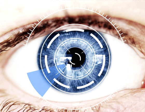
ALL in ONE the professional system for diagnosis of dry eye
With EASYTEAR® VIEW + you can easily visualize in vivo the interference fringes of the lipid layer in tear film, consider height of tear meniscus, with grids evaluate NIBUT (Non Invasive Break-Up time), evaluate BUT (Break-Up time), NIDUT (Non Invasive Dehydration Up time) to evaluate the wettability of contact lenses, observe and evaluate the Meibomian glands and finally evaluate the pupil diameter in various lighting conditions.
EASYTEAR® VIEW + is a complete instrument for tear film test (ALL in ONE) because it replaces the classics test lacrimal and reduces the sitting time, with obvious benefits.
EASYTEAR® VIEW + allows you to perform the main non invasive tear assessment tests aimed at diagnosis of dry eye. It through the analysis of the tear allows you to choose the best eye drops for target the root cause of dry, red, or itchy eyes without worsening existing irritation and choose the best contact lens specifications between many type of polymer in the world market.


THE FEATURES OF VIEW+
EasyTear® VIEW+ is very easy to use. It has a display and three intuitive buttons with simple icons for choosing the type of illumination and regulating across 5 levels with the help of an electronic control system. It has a seconds timer allowing calculation of average times for the specific analytic tests for blinking, NIBUT, BUT, NIDUT.


Operation of the ALL in ONE system and Dry Eye Disease (DED) Software
WHITE LEDS
The VIEW+ dacryoscope incorporates a series of white LEDs that combine with a special diffuser to generate wide corneal reflection for in vivo observation of the variable condition of the tear film and the anterior segment of the eye.
Lipid layer analysis
Definition of evaporative dry eye (EDE)
Observing the phenomenon of interference fringes enables calculation of the thickness of the lipid layer secreted by the Meibomian glands, quickly and precisely classifying it into 5 different categories (LLT). The non-invasive break-up time (NIBUT) can be assessed, with non-invasive in vivo observation of pre contact-lens film dry-up time directly on the eye (NIDUT). Quick and very easy image or video acquisition with HDWcam enables the operator to classify and quantify each individual condition according to international grading scales.
Tear meniscus
Definition of aqueous component deficiency (ADDE)
The aqueous layer on the tear meniscus is assessed, categorizing the possible associated problems into different species/classes/groups. The acquisition of multiple images enables direct noninvasive assessment of the aqueous tear content without reflex tearing.
Grids
The VIEW+ dacryoscope is supplied with a kit of 6 grids of different graphic designs to better highlight the drying of tears on the eye (NIBUT), pre contact-lens dehydration (NIDUT), and regularity of the corneal surface.
BLUE LEDS
The blue LEDs of the VIEW+ dacryoscope provide a wide zone of illumination over the entire anterior segment of the eye.
Fluorimetry
The VIEW+ dacryoscope incorporates six blue LEDs that, with fluorescein, enable a detailed analysis of the mucin layer and observation of conjunctival or corneal staining. With HDWcam, real time video becomes a fundamental tool for contact-lens opticians, revealing the movement of contact-lenses, the distribution of tear film below the lens, and the dehydration of the external face of the contact-lens. It is also possible to assess the tear film break-up time (BUT), making it possible to quantify tear stability and quality.
The DED analyzer software offers comparison of the observed conditions against an assessment scale.
INFRARED LEDS
Meibography
Thanks to the infrared illumination of VIEW+, the upper and lower eyelid Meibomian glands can be captured by HDWcam and analyzed by the DED analyzer software. The tarsal conjunctiva is the palpebral sector observed in order to best assess dysfunction of the Meibomian glands (MGD). This condition can cause or exacerbate dry eye symptoms and eyelid inflammation. The glands become blocked with solid secretions and if chronically clogged they ultimately become incapable of secreting lipids, inducing permanent changes in the tear film. With the ALL in ONE system, the Meibomian glands can easily be observed and compared with previous examinations of the patient/user to quantify the patients loss or their drop out (MGD). A practical MGD lever to facilitate eyelid eversion is provided as an accessory with the HDWcam device. The DED analyzer software allows comparison of the glands against a reference assessment scale.
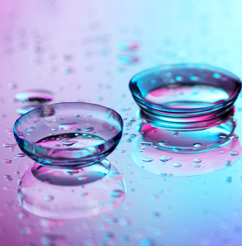
EASYTEAR® ALL in ONE – HELP TO REDUCE CONTACT LENS DROPOUT
The success of using contact-lenses depends on the comfort of the wearer, which in turn is closely linked to the function of the tear film. The identification and treatment of eyes with tear issues in contact-lens users, both new and established, is fundamental for successful implementation. In line with the specifications of TFOS DEWS, the ALL in ONE system by EasyTear® enables assessment of tear alterations with analysis of the lipid interference fringes, the tear meniscus, and the Meibomian glands. The contactlens specialist can use the collected information to choose the most appropriate polymer for the contact-lenses to best customize and satisfy individual customers. Furthermore, in routine checks on contact-lens users it is possible to use tear observations for early intervention in order to reduce drop-outs and sustain use of contact-lenses in complete safety.
EasyTear® VIEW+ offers a series of screening tests for dry eye syndrome
FUNCTIONALITY TESTED AND VALIDATED BY ITALIAN UNIVERSITIES
EasyTear® VIEW+ dacryoscope was tested at two Italian University to corroborate the effectiviness and functionality of the instrument:
The degree course in optics and optometry at the University of Milano-Bicocca validated the effectiveness of the white LED illumination system with precise levels of illumination to minimize the reflex tearing induced by examination. EasyTear® VIEW+ was found to be better than the original Tearscope® in terms of luminosity. It is capable of easily revealing the lipid layer interference fringes in the tear film. The validation included the blue LEDs, confirming the good diffusion features of thelight and wide field of observation of the slit lamp. The study was conducted by Dr. Luca Benzoni, Dr. Rossella Fonte, and Dr. Silvia Tavazzi who directed the project.
The University of Salento opearated at Lecce contact-lens resarch center under the scientific supervision of Prof. Giancarlo Montani to validate the infrared LED illumination system integrated into EasyTear® VIEW+. Ease of use for observing the Meibomian glands was compared with other existing devices.
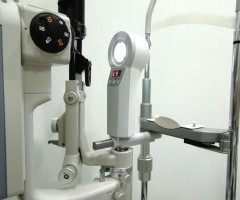
COMPATIBLE WITH ANY SLIT LAMP!
The EasyTear® VIEW+ instrument has a large through hole enablig binocular observation with the slit lamp.
Even the smallest details are better assessed in three dimensional vision. Considering the need for very close observation of very small details and varations in tearing, EasyTear® VIEW+ and HDWcam devices were designed for use on dedicated fixed stations or fitted on slit lamps of any type or brand with an universal bracket, however it is also possible to use them freehand.
The latter is very userful for example in cases when remaining still on the chin rest is difficult for certain patients during testing (e.g. the disabled, children, etc,), or when it is necessary to quickly check a contact-lens application without moving the patient from one work station to another, or during home visits.